Copyright 2012 neutronsources.org | All rights reserved. | Powered by FRM II | Imprint / Privacy Policy
Neutrons in Biology: Personal reminiscences of early days
By G. (Joe) Zaccai (CNRS IBS, ILL), 29/09/2014
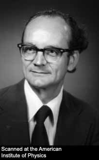
Grenoble, on summer holiday, was hot and quiet when I arrived at the ILL on August 1st 1974. It was long before the tram, and children played on the street in front of the cafés of avenue Alsace Lorraine. Previously at BNL on an EMBO fellowship, I had worked on model lipid membranes using Benno Schoenborn’s low angle diffractometer, and Mick Lomer, the ILL UK associate director, suggested I shared responsibility with Stewart Wilson to develop D16 as a membrane diffractometer. D16 was one of the two MK6 4-circle diffractometers imported from Harwell, which had been installed on a cold source beam in the guide hall. Bernard Jacrot had just returned from Cambridge where he had spent a year in Aaron Klug’s lab (Nobel Prize 1982), after his stint as first French director of the ILL.
I received my PhD in 1972 from the University of Edinburgh. My thesis project in solid-state physics included neutron diffraction on the Badger diffractometer at the DIDO reactor in Harwell. At the time, Harwell was opening up to outside research and Terry Willis was welcoming students and young scientists from universities to become neutron users. I met Sax Mason from Dorothy Hodgkin’s laboratory (Nobel Prize 1964), who was collecting anomalous dispersion and H-D exchange neutron crystallographic data on insulin. Also in the Hodgkin/Willis collaboration, Frank Moore and Brian O’Connor had solved the crystallographic structure of vitamin B12 using the automated Ferrante Mk1 neutron diffractometer built by Uli Arndt and Terry Willis at the PLUTO reactor, and Hartmut Fuess was working on neutron crystallography of insulin almost at the same time as the X-ray structure was being solved. It was my first contact with biological applications of neutrons. Neutron data collection required very large crystals and proceeded at a rate of a few reflections per day. In the same period, Walter Hamilton with Michel Frey, Jacques Verbist, Mogens Lehmann and Tom Koetzle at the chemistry department of BNL, and R. Chidambaram and his collaborators at BARC in India, were determining the crystal structures of amino acids by neutron diffraction to elucidate hydrogen bonds.
Similarly to structural biology being dominated by X-ray protein crystallography, early neutrons in biology studies were driven by neutron protein crystallography. In John Kendrew’s (Nobel prize 1962) laboratory in Cambridge, Benno had shown by X-ray crystallography how xenon atoms bound to specific sites in myoglobin and hemoglobin, studies that were published in Nature in 1965. These results had implications many years later when the xenon binding sites were used to analyze the molecular dynamics of ligand binding and release within the protein and its relation to the oxygen carrying function in myoglobin and hemoglobin. And recently, after the Sochi winter Olympics, I read a press report about suspicions that Russian athletes had boosted their oxygenation by breathing xenon gas, a practice prohibited by the World Anti-Doping Agency. At the biology department at BNL, Benno collected data at the HFBR on what were and still are considered to be enormous crystals of myoglobin to demonstrate the feasibility of neutron crystallography on proteins in a 1969 Nature publication.
In parallel with crystallography, a range of neutron applications in biology was being developed in the early 1970s. At BNL, Don Engelman and Peter Moore started SANS data collection on ribosomes on Benno’s low angle diffractometer, a project that took more than a decade to be accomplished. The complete mapping of the proteins in the 30s ribosomal subunit was published in 1987 in Science. Kent Blasie and myself used the same diffractometer for a study of model membranes (published in PNAS, 1975). Under the influence of Kissinger, Nixon’s national security advisor, the USA and Soviet Union entered an era of détente, which opened up areas of cooperation between the two countries, including in science. Igor Serdyuk from the Institute of Protein Research in Pustchino was invited to BNL to do SANS experiments also on ribosomes. He had developed a contrast variation method involving the parallel use of different radiation (X-rays, neutrons and light). Later, a time-of-flight SANS instrument was built by Ostanevich at the pulsed reactor in Dubna.
In Harwell, David Worcester modified a Badger diffractometer for membrane diffraction and SANS. He published fundamental results on lipid membrane structure in a book edited by Chapman in 1972. With John Pardon, he revealed the structure of the nucleosome particle of chromatin showing that the DNA was wrapped around the histone protein core. Chromatin is the DNA/protein strand of genetic material that condenses into chromosomes. The fundamental result was published in Nucleic Acids Research in 1977. At ILL, Konrad Ibel and Michel Roth built the small angle cameras, D11 and D17, on cold source guides. They established SANS as a powerful method in polymer science and biology. Ron Ghosh, who had also been working on his PhD at Harwell when I was there, developed the benchmark SANS data reduction programs still in use today.
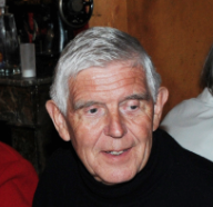
Heinrich Stuhrmann developed the mathematical formulation of contrast variation using H2O/D2O solvent exchange. Label triangulation was proposed independently by Engelman and Moore (in Proc. Natl. Acad. Sci (USA), 1972) and Walter Hoppe (in J Mol Biol, 1973). Engelman and Moore suggested using neutron scattering and specific deuterium labeling and applied the method to resolve the organization of proteins the 30S ribosomal subunit. At ILL, Knut Nierhaus, Roland May and collaborators, used label triangulation on the 50S ribosomal subunit and Hermann Heumann and his group also working with Roland May applied the method to solve subunit distances and shapes in DNA-dependent RNA polymerase. Water is essential for the processes of life, as we know it, to the extent that the search for extra terrestrial life is guided by the search for planets where water could exist in the liquid state. Water dynamics, in particular when it is confined in cells or other biological structures, is an important area of study, not only for biology, exobiology and origin of life studies but also in the context of medical imaging applications such as MRI and PET. And in what may well be the first report on biologically relevant molecular dynamics, John White measured the dynamics of confined water in clays, on the cold neutron spectrometer at DIDO (published in Nature, 1971). There is a powerful link between neutrons, water and biology link and during my first year at ILL a small group of us, with Peter Timmins, Tony Nunes and Mogens Lehmann, regularly went for ‘water’ lunches in the mountains to discuss possible experiments.
Most of the first instruments used in neutrons in biology were single detector diffractometers for chemical and physical crystallography. It was clear, however, that if neutron scattering were to become a viable method in structural biology a major instrumental effort would be required to counter the weak signal to noise from biological samples. At the HFBR, Benno built the dedicated low angle diffractometer using a 4.2 Å beam at the end of a short guide. Hoping to obtain a reduced background, the pyrolitic graphite monochromator was placed in an analyzer position, after the sample. Benno went on to inspire engineers in the instrumentation division of BNL (headed by Velko Radeka) to develop 2D position sensitive detectors. Large position sensitive detectors were developed and built at several of the reactor centers, with characteristics optimized for macromolecular crystallography, powder diffraction, small momentum transfer diffractometers or SANS. Variable focusing monochromators were designed and built to combine the advantages of large beam cross-sections, small samples and good angular resolution on position sensitive detectors. My first project during the EMBO fellowship at BNL was to work on an idea of Don Caspar to build a germanium/manganese multilayer and its bending support as a focusing monochromator for long wavelength neutrons, the ancestor of super-mirrors. D16, in the following decade, went from a ‘classical’ four circle diffractometer with a small single detector via steps including a large single detector and Soller slit collimation to enable the use of large samples, to the installation of progressively larger position sensitive detectors (as they became available) that could be scanned around the sample and a focusing monochromator with programmable vertical focusing. Neutron instruments for biological crystallography underwent a design revolution in the middle 1990s with the development of the quasi-Laue method driven by Mogens Lehmann and Clive Wilkinson and the development of imaging plates with neutron sensitive phosphors by Nobuo Niimura. Detector improvements to reduce effective dead-time led to SANS being able to cope with the large count-rates from biological samples. Biological dynamics studies, although still limited by the requirement for large samples, profited from major progress in instrumentation, culminating recently in the new IN5 and IN16B at ILL.
The ILL opened up neutron scattering to biophysicists, biochemists and molecular and cell biologists for the study of a range of problems in biochemistry and structural molecular biology. Previously, applications of the method were focused on a few well-defined problems, often using model biological systems to test methodological developments. The ILL proposal system made the facility widely accessible. The hiring of Peter Timmins, a chemist, and protein crystallographer with no previous neutron experience, stimulated studies to solve a variety of topical biological problems.
In 1973, after a successful career in neutron physics that led to his nomination as the first French director of ILL, Jacrot left to learn biology in Klug’s lab in Cambridge, where the new field of structural molecular biology was emerging. He shared a lab bench with Roger Kornberg (Nobel prize 2006), who was a young postdoc at the time working on chromatin. Jacrot worked on the biochemistry of viruses. His physicist colleagues mocked him. Normally, with his prestigious background, he would have been expected to take important responsibilities in French science. Yet, physicists contributed importantly to the development of molecular biology: Max Delbrück (Nobel Prize 1969) created the Phage Group in the early 1940s, an interdisciplinary network of scientists that unraveled bacterial genetics. Erwin Schrödinger published What is Life in 1944, a book that is still inspiring young physicists to become interested in biology. In the UK the physics-biology interface inspired by the appointment of Lawrence Bragg (Nobel Prize 1915) as Cavendish Professor at Cambridge in 1938 essentially led to the birth of a new field of science, structural molecular biology. Francis Crick and John Watson, and Rosalind Franklin and Maurice Wilkins were in physics departments, respectively, in Cambridge and at Kings College, London (headed by John Randall). Bill Cochran, who later was my PhD supervisor, had been in Cambridge at the time; with Crick, he calculated the Fourier Transform of a helix. Crick, Watson, and Wilkins were awarded the Nobel Prize in 1962. (Rosalind Franklin had died and the prize is not awarded posthumously). After his retirement, John Randall was active as an ILL user from the Zoology Department in Edinburgh.
Jacrot became Senior Scientist for Biology at ILL, then director of the Grenoble outstation of EMBL and went on to a senior position in the Life Sciences department of the CNRS. He taught me that to plan and perform a useful neutron scattering experiment in biology you had to understand the biological system and the question that was addressed. Sample preparation and characterization are paramount. I learnt to collaborate closely with biology users, often spending time in their lab to participate in sample preparation and to immerse myself in the biology of the problem in order to determine the appropriate neutron scattering protocol. And for my experiments on purple membranes on D16, I set up in the ILL basement a simple system to culture the organisms that produced the light sensitive membranes in a large heated balloon flask illuminated by fluorescent tubes. Biological systems are extremely complex making impossible a full characterization in terms of a physics model. A useful approach is to identify and measure appropriate physical parameters with respect to their relevance for biological function. The D16 results led to the fullest structural characterization of a natural biological membrane, and were published in a series of papers from 1979 in journals including J Mol Biol, PNAS, EMBO J, Mol Cell. The neutron diffraction work also paved the way for the first characterization of the thermal molecular dynamics of a natural membrane on IN13 at ILL, published in PNAS in 1993, following the work of Wolfgang Doster and his collaborators on the same instrument, describing the dynamical transition in myoglobin (published in Nature in 1989). Jacrot, working on D11, applied contrast variation to describe the nucleic acid and protein distributions in spherical viruses (published in Nature, 1977) —information that at the time could not be obtained reliably from either electron microscopy or X-ray diffraction. In a landmark study of nucleic acid-protein interactions using in-beam titration and contrast variation also on D11, Sylvain Blanquet, a biochemist, and Philippe Dessen, a biophysicist, deciphered the complexes formed and conformational changes in the interaction between tRNA and aminoacyl-tRNA synthetase, which ensures specificity of amino acid incorporation in the growing polypeptide chain during translation (published in J Mol Biol, 1978).
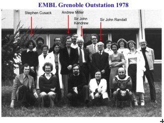
The EMBL outstation in Grenoble was established in 1976, in laboratories of the CENG across the fence from ILL. Andrew Miller was its first director, from 1976 to 1981, when the outstation moved on the site in a new building shared with ILL. The EMBL brought user-accessible state-of-the-art biochemistry and molecular biology to a few steps from the reactor and the site became a model for other neutron scattering centers. In the early eighties, Wally Koehler at ORNL and Mike Rowe and Jack Rush at NBS (later NIST) actively developed their respective centers for biology, incorporating in their plans the need for biology infrastructure dedicated to neutron users.
Disclaimer and apologies
The article is a personal account of the early days of neutrons in biology. It has no claim to being a properly researched historical document and I apologize in advance for errors due to a normally (so far at least) faulty memory.
