Copyright 2012 neutronsources.org | All rights reserved. | Powered by FRM II | Imprint / Privacy Policy
New Drug Carrier Aims to Treat Secondary Tumours of Breast Cancer
Date: 14/08/2015
Source: https://europeanspallationsource.se
COPENHAGEN — The battle to treat breast cancer—the most common cancer type for women globally—has spurred development of a new kind of drug delivery system for bone tumours associated with the disease. The system is based on bio-nanocomposites (bio-NCP), and is being developed through a collaboration of scientists from the Niels Bohr Institute (NBI) at Copenhagen University in Denmark, Universidade Estadual Paulista (UNESP) in Brazil, Helmholtz–Zentrum Berlin (HZB) in Germany, the ISIS neutron source and the University of Oxford in the UK, and the European Spallation Source (ESS) in Sweden.
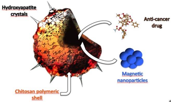
Previous studies have shown breast cancer cells migrate toward bone tissue due to the cells’ affinity for hydroxyapatite (HAP) [Ca10(PO4)6(OH)2], a primary mineral component of bone. Curiously, when reduced to the nanoscale, HAP is also a known inhibitor of cancer cell proliferation. Using this knowledge, the researchers designed a new bio-NCP drug carrier, which consists of an anti-cancer drug encapsulated in a polymeric nanosphere with a surface modified with HAP crystals. The crystals direct the carrier to the bone tissue and bind it selectively to cancer cells.
“The aim is to deliver the drug to the cancer cells in a selective and effective way with reduced pharmacological side effects,” says Murillo Longo Martins, the post-doc at NBI who led the study. “Magnetic nanoparticles were also included in the material, which allow us, for example, to track the position of the bio-NCP inside the body using magnetic resonance imaging.”
To decipher the chemical, physical and biological properties of the bio-NCP material and determine its effects on breast cancer cells, several state-of-the-art techniques were used, including thermal analysis, infrared spectroscopy, microscopy, preliminary biological tests, X-ray diffraction and neutron scattering. Neutron data was obtained with the FOCUS direct geometry spectrometer at the Paul Scherrer Institute (PSI) in Switzerland, the IN4 thermal neutron spectrometer at Institute Laue Langevin (ILL) in France, the FDS spectrometer at Los Alamos National Laboratory (LANL) in the US, and the POLARIS diffractometer at ISIS.
“The neutron powder diffraction data has shown how different chemical elements are distributed within the structure of the magnetic nanoparticles, which form the core of the bio-NCP. Neutron spectroscopy—inelastic and quasielastic—was used to study the molecular dynamics of an anti-cancer drug encapsulated into the bio-NCP,” says Heloisa Bordallo, who supervised Murillo at NBI, where she is an associate professor of X-ray and neutron science. Bordallo also serves as an advisor to the Scientific Activities Division at ESS. “The dynamical changes on this encapsulated molecule are currently being related to the biological properties of the bio-NCP against healthy and cancer cells.”
From a societal standpoint, development of the new bio-NCP holds promise to improve the efficacy and reduce the side effects of cancer drugs administered to patients.
Techniques applied:
- Thermal analysis: Differential scanning calorimetry (DSC) and thermogravimetric analysis (TGA) are considered to be among the most suitable approaches for determination of successful encapsulation and complexation processes. These analysis were are supplemented by FTIR to show that the loss of the melting peak of the pure drug is due to its complexation rather than thermal degradation or a loss of crystallinity.
- Infrared spectroscopy: An optimal tool to investigate the bio-NCP surface. The growth of the hydroxyapatite nanocrystals as well as the vibrational properties of the polymeric nano-sphere were investigated with this technique.
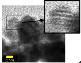
- Microscopy techniques: Scanning electron microscopy (SEM), transmission electron microscopy (TEM) and scanning transmission x-ray microscopy (STXM) have been used to provide insight on the morphology of the different components of the bio-NCP as well as the whole composite.
- X-rays and neutron diffraction: The most appropriate experimental tool for understanding the structure of the magnetic nanoparticles. Such information is the prior step to obtain particles with tunable properties for distinct applications.
- Inelastic neutron scattering: A unique tool to get insight into the dynamics of the drug confined inside the bio-NCP. Observing the dynamical behavior of the encapsulated drug was only possible thanks to the high penetration of neutrons into matter.
- Biological trials: In-vivo tests have been already performed in healthy and cancer cells and the biological properties of the bio-NCP are currently being evaluated.
Further Reading:

